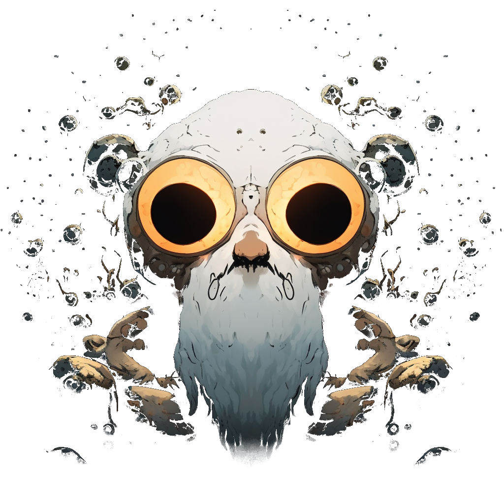
This is a photograph of a small trichome on the surface of a seedling through the 40x objective. Not sure if it is a happy trichome looking up at what it will become or a sad trichome looking down 😆 I liked the colors and the scene, reminds me of a painting.
Here is a photo through the 10x:

Seeing these posts kinda makes me want to buy a microscope. To research the poo of the pigeons on my balcony 😆
What kind of a microscope are you using? Do you do that just as a hobby?EDIT: I just checked and I actually have the SW380T, not 350T. It is similar but slightly better from what I can find: https://www.microbehunter.com/swift-sw380t/
Interesting! I ran a quick search to look into what you might see. I found a paper titled "ESTABLISHING NORMAL FECAL FLORA IN WILD AUSTRALIAN PASSERINE BIRDS BY USE OF THE FECAL GRAM STAIN, and in that paper they find no parasites in the feces sampled and mostly gram-positive bacterial with cocci shape. Yeasts were found some rarely. To actually identify the bacteria they use DNA sequencing. Other than the bacteria, if the sample is fresh, you might be able to find crystals (like uric acid), and epithelial cells… If the sample is a bit older you would also be able to identify microorganisms that feed on it.
The version I have at home is a SWIFT SW350T. It costs ~360 € in the EU. It has the eye pieces and third a tube for mounting a camera. For mounting the camera I purchased an adapter separately for my Nikon camera (it mounts to the DSLR camera as a regular lens and then the tube is inserted through the top port of the microscope). The SW350 (without ‘T’) is slightly cheaper at $300 and it missing the top port, but it is still possible to mount a camera through an eye piece. Having the third one is just a bit more convenient as you can look through the microscope while the camera is mounted.
I use it as a hobby. I have worked with a few fancy microscope types in a research setting, but then the kinds of samples I have looked at have been very restricted.
Ha, i was just thinking about the pigeon poo because it was what i was looking at the last time i looked through a microscope. And there were in deed also cocci bacteria there.
That looks like a real cool microscope, and not even that crazy expensive. And with an actual camera mount. I’ll have to let the thought simmer a bit, not just buy one to look through it a few times then have it collect dust. Gonna follow your microscopic adventures :) Thanks for the info!
I just noticed that the one I have is 380T, not 350T! The 380T is slightly more expensive but allegedly has a bit better of a view on the edges. Here is a review from microbe hunter: https://www.microbehunter.com/swift-sw380t/
It comes with a dust cover, so no worries ;)
I did let mine sit for a while at first. There is a learning curve to getting things on the plane and focused with good lighting, and the first several times using the microscope was a whole activity in itself, so I would only use for a special occasion when I wanted to spend time learning. Over time it becomes easier to prepare samples, keep the optics clean, and to know what you are looking at. The microscope becomes a useful tool rather than an activity, and then it is less likely to collect dust because the opportunities to use it become plenty.
Not trying to bias you though, take your time! Haha
It comes with a dust cover, so no worries ;)
Oh well, haha! I watched microbehunters review, looks like a real cool microscope. Are you using any other lighting than the integrated led?
And the camera adapter, this is not just a plastic (or metal) part to fit into the tube so the camera would use the actual microscope lens, but it’s something that has its own optics too?
I am using the integrated LED. Sometimes I use a UV flashlight to see fluorescence, and I would like to play with others. I work with spectroscopy and I have the intention of adding a bit of spectroscopy to the microscope, but I am still not there yet.
The camera adapter that I am using does contain some optics inside and has 2X magnification. It is one like this one: https://nl.aliexpress.com/item/32943613016.html
Microbehunter has a review on these: https://www.youtube.com/watch?v=Ju8rgeJr3bI
Roughly, if you use a 40x objective, the 2x adapter, and the camera is a DSLR with an APS-C sensor like mine (dimensions 23.6 mm x 15.7 mm), then the image that you capture with the camera corresponds to approximately the sensor dimensions / (objective multiplier x adapter multiplier) = 295 micron x 196 micron in this case. In practice it is good to use a microscope ruler for accurate dimensions,
There are camera adapter designs with no optics that you can 3D print, but I have not tested those. What I have tested are tubes that you can 3D print that connect the camera to an objective directly without the need for a microscope. This is can be useful for macro photography but the microscope is a lot more comfortable to use for general microscopy. This is what I mean: https://www.youtube.com/watch?v=nuZ_JptlqYE
This seems to be a whole field of rabbit holes. Tried to wrap my head around your calculation yesterday evening, and what it’d mean for my ancient m43 cameras, that was a bit confusing at first (where is the crop factor!), i have never really messed with by-x lenses. But i think the penny has eventually dropped, haha. Then i got sidetracked on how microscope rulers are being made. I reckon you have your setup calibrated.
Thank you for the interesting pointers.
that was a bit confusing at first (where is the crop factor!), i have never really messed with by-x lenses
Haha, I think I have a good idea of why… When thinking about general photography, common scenes involve many different types of objects of varying sizes and distances. In this context it makes sense to define the “field of view” of a lens in terms of the visual angle, and to think about differences between equipment in terms of a “reference” - that’s where the ‘crop factor’ comes in, when comparing to a 35 mm film as a standard to measure relative to. It is a bit silly to ask a question such as “how many cats can fit into the image that gets projected into the camera sensor?”
When one gets into macro photography things start changing. The distances between the camera and the subjects are defined more narrowly and the size of the subjects one tries to capture is closer in size to the camera sensor’s size. In macro-photography you hear about things like a “1:1” lens, meaning that the image of an object placed at a specific distance will be replicated 1:1 at the camera sensor’s position. It becomes then meaningful to think about the camera sensor size in absolute sense, because a sensor will capture a scene of its own size (for 1:1). You can still think in relative terms using the crop factor, but it is not as useful to make relative comparisons anymore because the absolute scale is already giving us information that we can work with directly.
For microscopy this trend continues - the distance to the subject is well-defined and we can think in absolute terms about the magnification of the image that gets projected into camera sensor.
Then i got sidetracked on how microscope rulers are being made.
I have not looked into this! I am not sure… I will look it up.
I reckon you have your setup calibrated.
I do have a ruler but I misplaced it and I have not used it in a while. So, not really, I wouldn’t say it is “calibrated”. I did take some photos of the ruler that I can use to get a good rough estimate if I can find them.